

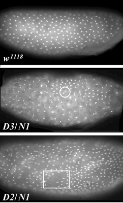
The image at right shows three Drosophila embryos at the syncytial blastoderm stage of development. DNA is stained with the dye DAPI. At this stage, nuclei are present in a syncytium, with no cell membranes between them. The cell cycle is extremely rapid, consisting only of S and M phases, with a mitosis every 6-8 minutes.
The top embryo is from a wild-type mother (she was actually w1118, but wild-type for mus309. Note the evenly ordered distribution of nuclei.
The middle embryo is from a mother that lacks DmBLM because her genotype is mus309D3/mus309N1. Two defects are apparent. First, the nuclei are asynchronous: Some are clearly in interphase (S), whereas others appear to be in anaphase. Second, there are anaphase bridges between DNA masses (circled).
The bottom embryo is from another mother that is mutant for mus309, but in this case mus309D2 (a nonsense mutation) replaces mus309D3 (a missense mutation that changes the DEAH motif to DKAH); embryos from mothers of these two genotypes are not distinguishable. This particular embryo is one or two cell cycles more developed than the middle embryo. This image was chosen to highlight another aspect of the mutant phenotype: gaps in the normally uniform monolayer of nuclei (square). This happens when nuclei are defective and a system that detects such nuclei causes them to sink in to the middle of the embryo, where they will become part of the yolk and not contribute to the tissue of the larva. This won't matter in this case, because these embryos would not survive to the larval stage, even if we hadn't fixed and stained them.
This phenotype was described inM. McVey, S. Andersen, Y Broze, and J. Sekelsky (2007) Multiple functions of the Drosophila Blm helicase in maintaining genome stability. Genetics 176: 1979-1992.