

|
This image of a Drosophila female meiotic nucleus was made by Abby Dernburg. DNA was labeled with DAPI, and flourescence in situ hybridization (FISH) was done to detect sequences near the centromere of each chromosome: | 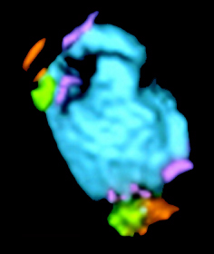
|
|
|
| Ā | |
||||||||
|
|
© Newtron Laboratories